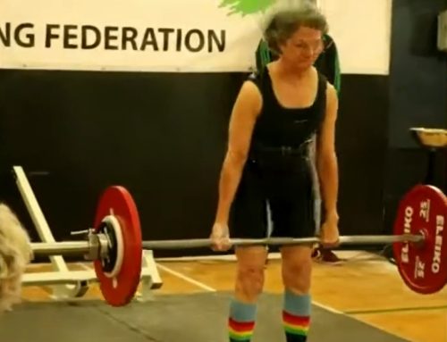

 To become a Champion of Aging, you must train to build strength and muscle.
With that in mind, it is helpful to understand the anatomy of our muscles to gain more confidence in our approach to training.
In order to look at muscle anatomy, we’ll need to get a bit into the science weeds. Much of what follows was derived from my online training at Barbell Logic.
However, this in depth look will provide you with a better understanding of why it is necessary to employ different training modalities to become a Champion of Aging.
Before I get into that, if you are interested in building out your own home gym, be sure you check out my Recommended Equipment once you finish this article.
Muscle Origin and Insertion
Every point where a muscle connects to bone, usually via a tendon, is an attachment.
Although muscles can take many shapes and often have many attachments, it is useful to think of most muscles by their origin and insertion.
Here are some general rules for understanding origin and insertion…
- The origin is usually on the more stable bone.
- In a two-joint muscle, the origin is almost always on the joint closest to the trunk.
- The insertion of a muscle is usually at its more moveable end. So, in this case, the insertion of the biceps brachii is on the radial tuberosity of the radius.
- The insertion moves toward the origin in the vast majority of cases.
Muscle Function and Size
Once you know where it attaches, it’s useful to consider the role it plays in movement.
A muscle may act as an agonist, antagonist, stabilizer, assister, or neutralizer in one movement, then take on a different role in a different movement.
In virtually every joint in the body, different muscles will play a role in the movement of that joint.
However, some will play a more dominant role and others will merely provide assistance.
In general, the size of the muscle and its location will determine whether it plays a more dominant role or not.
Muscle Fiber Direction
Like how a circle makes a better wheel than a square, a muscle’s shape and fiber direction make it more (or less) suited to different jobs.
Remember that the fibers of the muscle only shorten.

The layers and layers of them shaped in certain patterns affect both the amount of force that the muscle can produce and how it will act on a joint.
Many bigger muscles will have muscle fibers pointing in many different directions. This allows the muscles to do several seemingly opposite actions in different movements.
Additionally, a muscle’s fiber direction determines how far away that fiber is from the joint when it pulls.
This affects its moment arm so two muscles of the same size with the same general function can provide different amounts of force to a particular movement.
Basic Muscle Fiber Structure
 There are three muscle types in the human body: cardiac, smooth, and skeletal muscle, but for this course, we’ll focus on skeletal muscle.
When you see a muscle, you’re looking at a bundle of bundles: long fibers held together by wrappings of connective tissue called fascia.
The smallest working unit of the muscle is the myofibril. The myofibril is a long tube held together by fascia called endomysium. The myofibril is made up of sequential discs called sarcomeres that do the work of contraction.
A bundle of myofibrils is a myofiber, more typically known as a muscle fiber, and is held together by fascia called the perimysium. The nerves that cause muscle to contract interact with the sarcolemma, the outer membrane of a muscle fiber, and every myofibril in the muscle fiber responds at the same time.
A collection of muscle fibers bound by epimysium forms a whole muscle. Muscle causes movement by contraction: attempting to shrink towards its center, which creates a pulling force at all of its attachments.
Muscle Actions
Concentric action is when the muscle attachments move closer together in resistance to a load.
Isometric action is muscular action without the tissue changing length. This happens when clenching or flexing (like in a fixed bodybuilding pose) or pressing against an unmoving object.
Eccentric action is when muscular attachments are moving farther apart while the muscle fibers are trying to pull closer.
Muscle Fiber Types
To meet a broad range of force and energy demands, the body has two major fiber types:
- Type I: “Slow Twitch”
- Type II: “Fast Twitch,” divided into two subtypes, IIa and IIx.

Sprinters use more Type II fibers
Slow-twitch fibers are the smallest in diameter and are less energy-intensive.
They are so efficient because they have higher concentrations of mitochondria, myoglobin, and higher capillary density—allowing them to use oxygen most efficiently.
These fibers are referred to as aerobic for this high oxidative capacity.
As you might expect, because they are the smallest, they have the lowest force production, though they do not fatigue easily.
They are not the most excitable either, hence the slow-twitch name.
They have the slowest contraction times, which means they are slower to get up to their peak tension.
This comes in handy for long-term, low-energy activities like holding our posture in a yoga pose.
These fibers are present in every muscle and are recruited first, whether the lifter is running a marathon or completing a maximal deadlift.
Fast-twitch fibers are divided into two groups: type IIa and type IIx.
Both are bigger in diameter than type I fibers and, therefore, can produce greater force more quickly.
Type IIx is the quickest and strongest but also the most energy-intensive.
The trade-off for this power is fewer mitochondria, myoglobin, and capillary density (type IIx having the least of all three).
That makes these fibers fatigue more quickly because they are not efficient at generating ATP aerobically, which they need for sustained output.
Muscle Fibers and Training
 Every skeletal muscle has all three fiber types in different proportions.
Muscles that are responsible for sustained low-intensity force (like holding a sitting posture) tend to be predominantly Type I.
Peripheral muscles that typically produce great force tend to have higher proportions of Type II fibers.
This is also influenced by genetics. Some people appear to be born with higher proportions of Type I or Type II muscle fibers.
However, even a muscle fiber’s type is trainable to some extent.
For now, remember that adaptations to training can be as obvious as bigger muscles or as subtle as fine-tuning your muscle fibers to behave one way or another.
From Brain to Muscle
When you voluntarily move a muscle, this is the general pattern of how that signal gets from the brain to the muscle:
- An electrical signal starts at an upper motor neuron (UMN) in the motor cortex of the brain.
- That upper motor neuron sends an electrochemical signal along its axon to a lower motor neuron (LMN).
- Most lower motor neurons are in the spinal column—though a few, like those that control the muscles of the eye, are distinct.
- The signal excites the LMN associated with the target skeletal muscles.
- The LMN sends an electrical signal along its axons to every muscle fiber that it connects to.
- The electrical signal reaches the neuromuscular junction (NMJ), which is the point where the axon terminal meets the muscle fiber and triggers the release of acetylcholine (ACh), a neurotransmitter.
- At the junction between the axon and the muscle is a gap, a synapse.
- Acetylcholine crosses the synapse and binds to receptors on the sarcolemma of the muscle fiber to trigger another electric signal.
- The electrical signal at the muscle side of the NMJ propagates across the sarcolemma and enters the muscle through channels called T-Tubules, which releases calcium ions into the cytoplasm of the muscle cell.
From muscle to movement
The long, thin tube-shape of the muscle cell is divided into compartments called sarcomeres.
Each sarcomere is bounded on each side by a z-disc: a flexible wall that anchors an actin filament.
Myosin uses hook-like heads to attach to the actin. At rest, myosin is blocked from the next available location along the actin by another protein called troponin.
When calcium enters the cell, the troponin changes its shape, exposing the next available ‘hook’ on the actin for the myosin to grab onto.
ATP (Adenosine Triphosphate) is used to release the myosin from its resting position (‘cocked’), where it returns to its natural extended position, bringing it farther along the actin where it hooks and curls, pulling the actin towards the center of the sarcomere—ready to repeat the process with new ATP.
As the actin is pulled inward, so is the z-disc the actin is anchored to, and the entire sarcomere literally shrinks as its walls are pulled toward the center.
Repeated along thousands of sarcomeres, and within all the muscle fibers in a motor unit, this squeezes the muscle inward on itself, creating a pulling force at the ends of the muscle where it attaches to bone.
Final Thoughts
We previously covered how the body adapts to strength training.
This deep dive into muscle anatomy provides a bit more background in regard to why our muscles adapt to different types of training.
It’s not necessary for a trainee to have a complete understanding of anatomy and physiology.
However, I’ve found that many people tend to default to easier exercise routines as a result of a lack of knowledge of the science behind the training.
As a result, they really don’t train hard enough of consistently enough to achieve the results they want.
To learn more about strength training in general, check out this article on the Principles of Strength Training.